Let us create a wonderful future together!
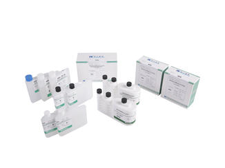
An Amyloid A Assay Kit detects serum amyloid A in human serum in vitro. This test is an excellent nonspecific inflammation indicator, as the level of amyloid A protein increases sharply during inflammatory, infectious, or non-infectious conditions. This protein is also present in the blood of people suffering from diabetes, atherosclerosis, or chronic kidney disease. Amyloid A Assay Kits are available from a number of manufacturers.
Amyloid A is an acute phase reaction protein
Acute-phase reactions are characterized by an increase in plasma proteins. These changes begin within hours or days of the onset of most tissue damage and continue for several days or weeks. The main acute-phase reactants are serum amyloid, coagulation factors, and transport proteins. The minor acute-phase reactants include fibrinogen, haptoglobin, and ceruloplasmin. Each of these proteins has its own biological and clinical value.
In the serum, acute-phase SAA proteins are multifunctional apolipoproteins that play a role in cholesterol transport, metabolism, and immunological responses. These proteins are induced by pro-inflammatory cytokines and are capable of increasing in concentrations up to 1,000-fold. A chronic elevation in serum amyloid A proteins is a prerequisite for secondary amyloidosis, a fatal and progressive disease in which the deposition of insoluble plaques in the tissues are the primary symptom.
The body produces these proteins to strengthen its innate defenses against infection. However, too much production can lead to serious problems and even shock. In addition, some APRs may have deleterious effects on health. Amyloid A is an example of an acute phase reaction protein. Once again, this article will discuss the types of APPs and the role they play in vaccines. Once you understand more about APRs, you will be better equipped to decide how to use them in the future.
It is detected by ELISA
The ELISA test for serum amyloid A (SAA) measures the level of this acute-phase protein in human serum. The ELISA kit uses a strip-well format to measure the level of amyloid A in human serum. The kit contains reagents for as many as 96 tests. Amyloid A is associated with rheumatoid arthritis and sarcoidosis.
The ELISA protocol for detection of Amyloid A has been validated by several studies. ELISA measurements below 110 pg/mL did not have any correlation with those from SIMOA. In addition, the Ab1-40 measurements had a floor effect that caused the data points to deviate from the Passing-Bablok regression curve. This poor commutability is evident in Bland-Altman plots.
Currently, two types of ELISAs are available for detecting amyloid A in human serum. One is a paper-based ELISA, while the other uses a 96-well format. The ELISA used by Millipore detects Amyloid b40 in cerebrospinal fluid. The assay is highly sensitive and has a sensitivity of more than 96%.
Commercial ELISA kits for Amyloid A are available from EUROIMMUN in Lubeck, Germany. Prototype SIMOA Amyblood assays are also available. Both use the same monoclonal antibodies, which bind to residues 1-4 of the Ab peptide. During the tests, TMB is directly proportional to the concentration of o-Ab. This method allows for quick and accurate determination of levels in human urine.
It is used to quantify levels of serum amyloid A
The method used for the measurement of serum amyloid A (SAA) is a highly sensitive and specific test. The assay requires six-uL of human serum or plasma as a standard sample. The sensitivity of this test is 0.5 ng/mL with a dynamic range of 1.56-100 ng/mL. It is manufactured by Crystal Chem, a world-renowned manufacturer of ELISA kits.
Although its direct role in procoagulation is not clear, serum amyloid A is a useful biomarker for coronary artery disease. One study studied the relationship between serum amyloid A and tissue factor pathway inhibitor. It is now regarded as an independent biomarker for coronary artery disease. However, despite the evidence for this link, no large-scale clinical studies have been performed.
This test uses anti-SAA antibodies to detect serum amyloid A levels. The cells stained with the anti-SAA antibodies have high levels of SAA. Cells stained with SAA exhibit brown staining. In RA tissue, the protein is expressed by synovial vascular endothelial cells and perivascular areas. In OA, however, SAA expression is low. The SM contains fibroblast-like synovial cells.


